Puccinia graminis tritici
Stages on the primary host; Wheat
1. Uredospores (on wheat)
- Uredospores are borne in groups under the epidermis, called uredosorus.
- The uredosorus develops on wheat plant from the dikaryotic mycelium produced by germination of aeciospore.
- The uredospores appear in the form of reddish-brown pustules.
- The uredospores are stalked, oval, unicellular, brown, thick walled with 4-round equatorial germ pores.
- The spore wall is thick with the echinulate outer layer.
 |
Uredosorus showing stalked unicelled, binucleate uredospores
|
2. Teliospores (on wheat)
- Teleutosorus develops exclusively by the infection of uredospore.
- The teleutosori look black raised streak on leaf sheath and also on stem.
- The teleutospores are stalked, spindle-shaped, thick and smooth-walled with round or pointed apex, 2-celled and slightly constricted at the septum.
- Spores are chestnut brown in color.
- Each cell is dikaryotic (n + n) having one germ pore.The germ pore is at the top of the apical cell and it is below the septum in the lower cell.
 |
Teliosorus showing stalked bicelled, binucleate teliospores
|
|
Stages on the primary host; Barberry
3. Pycnidiospores (on barberry)
- Pycnidium is developed under the upper epidermis in the form of a yellowish flask-shaped structure.
- The pycnidium has small sterile mycelium at the neck, called periphyses, which intermingle with much larger thin-walled simple and branched receptive or flexuous hyphae.
- The bottom of the inner side of pycnidium is lined by many uninucleate tapering cells, the spermatiophores, which develop many small oval to spherical uninucleate cells, called pycniospores (spermatia).
- The pycnidiospore may be of + or – type.
- The mature pycnidium (+ and – type) secretes nectar drops during release of mature pycnidiospores which get intermixed.
- The insects get attracted by nectar and help in the transfer of pycnidiospores to the flexuous hyphae of opposite mating type.
- The wall at the point of contact dissolves and the nucleus of the pycnidiospore passes to the flexuous hyphae, thus a dikaryotic condition is established. This process is known as spermatisation.
 |
| Pycnidial cup showing the pycnidiospores |
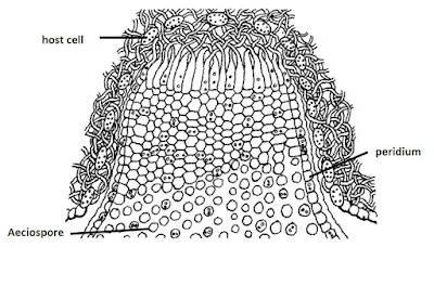
4. Aeciospores (on barberry)
- The young aecium is borne inside the tissue below the lower epidermis, but with maturity it pushes and ruptures the epidermis, thereby spores are exposed.
- liThe aecium is an inverted cup-shaped structure with outer margin composed of short cells, called peridium.
- The stalk cell after becomes dikaryotized, divided mitotically into a chain of alternately arranged large and small cells.
- The large cells form the aeciospores (n + n) and the small one becomes sterile, called the disjunctor cell.The disjunctor cell helps in spore dispersal.
- The aeciospores are unicellular, binucleate (n + n), thin-walled and orange in colour.
- The young aeciospores are polyhedral in shape, but become globose with maturity.
 |
| Aecial cup showing chain of unicelled, binucleate aeciospores |
Content first created on 01-09-2023
last updated on 01-09-2023


0 Comments
Leave your comments here.